Figure 3.
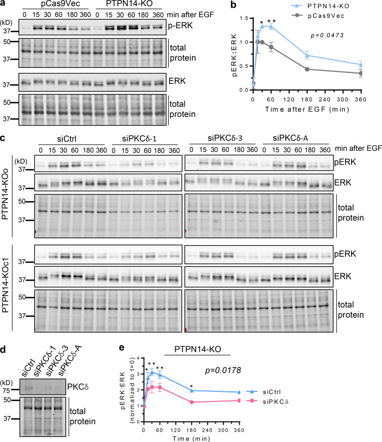
pY374-PKCδ enhances ERK activation downstream of EGFR. (a and b) ERK activation after stimulation with EGF (20 ng/ml), for the indicated times, was analyzed by Western blotting with rabbit anti-phospho-ERK and mouse anti-ERK Abs in pCas9Vec and PTPN14-KO cells (a) and quantified (b; mean ± SD from n = 2 pCas9Vec and n = 2 PTPN14-KO clones, P = 0.0473 using two-way ANOVA followed by Sidak’s multiple comparisons test for each time point; *, P < 0.05; **, P < 0.01). (c) PTPN14-KO cells transiently transfected with control (siCtrl) or three different PKCδ (siPKCδ) siRNAs with phospho-ERK and total ERK Western blots shown. (d and e) PKCδ blots (d) and ratio of pERK to total ERK blots quantified (e; n = 2 PTPN14-KO clones, each transfected with siCtrl and three different PKCδ siRNAs (averaged for each clone); P = 0.0178 using two-way ANOVA followed by Sidak’s multiple comparisons test for each time point; *, P < 0.05; **, P < 0.01).

