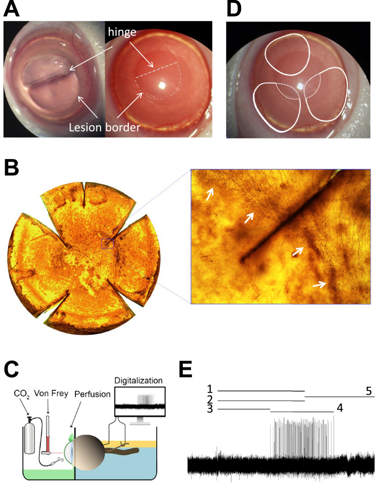
Figure 1.
(A) Guinea pig cornea showing the corneal flap, the border of the lesion and the hinge area, immediately after surgery. Arrows point the lesion border and the flap hinge (dashed line). (B) Flat-mounted cornea stained with Tuj-1 displaying the corneal flap absence of nerves inside the flap and the presence of Tuj1-positive subbasal nerve leashes outside the lesion. The inset shows how nerves bench at the border lesion without entering the flap. Arrows point the lesion border. (C) Schema of the ciliary nerve recording set-up. (D) Representation of the receptive field (RF) of three different nociceptive fibers. One is located at the peripheral cornea and the hinge area (up), that is, outside the lesion. The other two RFs covered the peripheral cornea (continuous lines) and presumably also part field inside the wound (dashed lines), but no activity was recorded when stimulating inside the lesion at any time point. (E) Sample recording of the nerve activity evoked in a polymodal nociceptor in response to CO2 pulse, showing the parameters analyzed to characterize chemical responsiveness: 1 is the duration of the CO2 pulse; 2 is the nerve activity during the pulse; 3 is the latency to the response; 4 is the response; 5 is the postdischarge (see Methods for further details).

