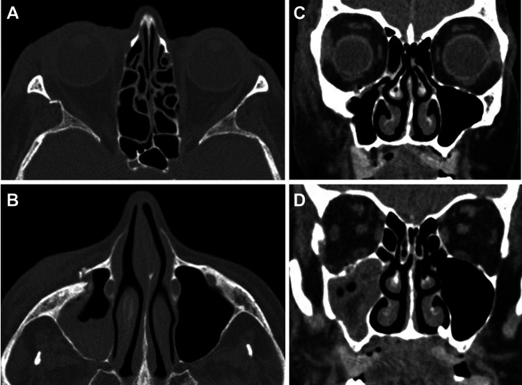
Figure 1.
Preoperative CT scans demonstrating: (A) axial section with right lateral orbital wall fracture with free-floating segment, (B) axial section with right displaced inferior orbital rim, (C) coronal section with depressed fracture of the lateral wall of the maxillary sinus, and (D) coronal section showing posterior lateral maxillary sinus fracture, lateral orbital wall fracture, and small orbital floor fracture adjacent to the infraorbital canal. CT indicates computed tomography.

