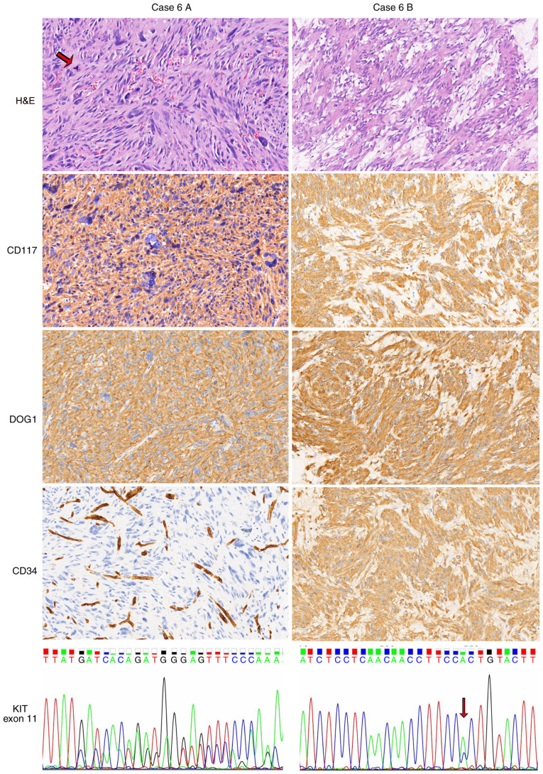
Figure 3.
Heterogeneity of GISTs in a second patient (case 6). Hypercellular and pleomorphic histology was visible in the specimen of case 6A, a gastric GIST. Multinucleated giant cells and pathological mitoses (arrow) were observed. The tumor in case 6A was positive for CD117 and DOG1 but negative for CD34. Endothelial cells served as an internal positive control. Case 6B, a GIST between the stomach and spleen, exhibited a conventional spindle cell morphology, with the presence of myxoid stroma and the strong expression of CD117, DOG1 and CD34. KIT codon 579 was deleted in case 6A, whereas a KIT exon 11 substitution (p.W557G, arrow) existed in case 6B. Images show H&E, CD117, DOG1 and CD34 staining (magnification, ×200). GIST, gastrointestinal stromal tumor; DOG1, discovered on GIST-1; KIT, KIT proto-oncogene, receptor tyrosine kinase; H&E, hematoxylin and eosin.

