. 2021 Jan 7;3(1):e200535. doi: 10.1148/ryct.2020200535

Figure 1c:

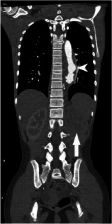
(A) Chest radiograph shows cardiomegaly and an abnormal left paraspinal density in the region of the descending thoracic aorta (arrow). (B, C and D) Coronal contrast-enhanced computed tomographic angiography images of the chest, abdomen and pelvis reveal a diffuse abnormal appearance of the descending thoracic aorta and suprarenal abdominal aorta with lumen irregularity, wall thickening and/or mural thrombus as well as multiple aneurysms (arrowheads). Lack of contrast opacification of the left renal artery and delayed enhancement of the left kidney (white arrow) secondary to renal artery stenosis or thrombosis.

(A) Chest radiograph shows cardiomegaly and an abnormal left paraspinal density in the region of the descending thoracic aorta (arrow). (B, C and D) Coronal contrast-enhanced computed tomographic angiography images of the chest, abdomen and pelvis reveal a diffuse abnormal appearance of the descending thoracic aorta and suprarenal abdominal aorta with lumen irregularity, wall thickening and/or mural thrombus as well as multiple aneurysms (arrowheads). Lack of contrast opacification of the left renal artery and delayed enhancement of the left kidney (white arrow) secondary to renal artery stenosis or thrombosis.