. 2021 Jan 7;3(1):e200535. doi: 10.1148/ryct.2020200535

Figure 2b:

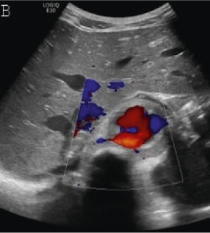
(A,B,C,D) Transverse and sagittal gray-scale and color Doppler ultrasound images of the upper abdominal aorta (white arrow) and (E,F) transverse gray-scale and color Doppler ultrasound images of the superior mesenteric artery (black arrow) show wall thickening and/or mural thrombus, lumen irregularity, and multiple aneurysms. (G,H) Transverse Gray-scale and color Doppler ultrasound images of the abdominal aorta and left renal artery (yellow arrow) show ostial narrowing of the left renal artery.

(A,B,C,D) Transverse and sagittal gray-scale and color Doppler ultrasound images of the upper abdominal aorta (white arrow) and (E,F) transverse gray-scale and color Doppler ultrasound images of the superior mesenteric artery (black arrow) show wall thickening and/or mural thrombus, lumen irregularity, and multiple aneurysms. (G,H) Transverse Gray-scale and color Doppler ultrasound images of the abdominal aorta and left renal artery (yellow arrow) show ostial narrowing of the left renal artery.