. 2021 Jan 7;3(1):e200535. doi: 10.1148/ryct.2020200535

Figure 3c:

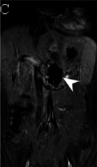
(A) Axial triggered angiography non-contrast enhanced (TRANCE) MRI sequence shows suprarenal abdominal aortic aneurysm (star) and marked decreased signal intensity of the left renal parenchyma (arrow) compared to the right consistent with renal ischemia secondary to decreased perfusion. (B) Axial echo-planar image (EPI) MRI image and (C) Coronal EPI post-contrast MRI image reveal non-enhancing aortic wall thickening (black arrow head) suggesting wall edema and/or mural thrombus. There is also undulating contour of the aortic lumen with saccular dilatation (white arrow head). (D) 3-D reconstructed image reveals the luminal irregularity and aneurysms of the thoraco-abdominal aorta.

(A) Axial triggered angiography non-contrast enhanced (TRANCE) MRI sequence shows suprarenal abdominal aortic aneurysm (star) and marked decreased signal intensity of the left renal parenchyma (arrow) compared to the right consistent with renal ischemia secondary to decreased perfusion. (B) Axial echo-planar image (EPI) MRI image and (C) Coronal EPI post-contrast MRI image reveal non-enhancing aortic wall thickening (black arrow head) suggesting wall edema and/or mural thrombus. There is also undulating contour of the aortic lumen with saccular dilatation (white arrow head). (D) 3-D reconstructed image reveals the luminal irregularity and aneurysms of the thoraco-abdominal aorta.