. 2021 Jan 7;3(1):e200535. doi: 10.1148/ryct.2020200535

Figure 4:

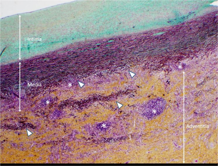
Histopathological slide with Movat stain, 20x magnification. Noninfectious aortitis with patchy perivascular, adventitial and medial lymphocytic infiltrates (*). There is moderate thickening of the intima, variable medial thickening and disruption of elastic media (Δ) and marked, dense fibrotic thickening of the adventitia.

Histopathological slide with Movat stain, 20x magnification. Noninfectious aortitis with patchy perivascular, adventitial and medial lymphocytic infiltrates (*). There is moderate thickening of the intima, variable medial thickening and disruption of elastic media (Δ) and marked, dense fibrotic thickening of the adventitia.