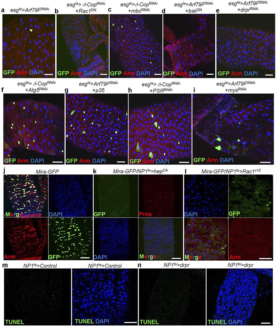
Extended Data Figure 8 |. Knockdown of components of the JNK pathway or engulfment genes in ISCs did not block the ISC death induced by Arf79FRNAi or δ-CopRNAi expression.

a–i, The genotypes of the flies in each panel were: a, esgts > Arf79FRNAi, 29 °C, 7 d (n = 27). b, esgts > δ-CopRNAi + Rac1DN, 29 °C, 7 d (n = 32). c, esgts > δ-CopRNAi + mbcRNAi, 29 °C, 7 d (n = 25). d, esgts > Arf79FRNAi + bskDN, 29 °C, 7 d (n = 30). e, esgts > Arf79FRNAi + drprRNAi, 29 °C, 7 d (n = 28). f, esgts > δ-CopRNAi + Atg5RNAi, 29 °C, 7 d (n = 32). g, esgts > Arf79FRNAi + p35, 29 °C, 7 d (n = 22). h, esgts > δ-CopRNAi + PSRRNAi, 29 °C, 7 d (n = 30). i, esgts > Arf79FRNAi + mysRNAi, 29 °C, 7 d (n = 28). bskDN is a dominant-negative form of Drosophila JNK (ref. 51), draper (drpr) encodes a homologue of the C. elegans transmembrane phagocytic receptor (ref. 52), Rac1 encodes a small GTPase that is a homologue of the C. elegans engulfment gene ced-10 (ref. 53), myoblast city (mbc)/Crk/dCed-12 encodes a Rac1 guanine nucleotide exchange factor (GEF) (ref. 53), PSR encodes a phosphatidylserine receptor (ref. 54) and mys encodes the β-subunit of integrin, which is involved in mammalian cell engulfment (ref. 55). Light chain 3 (LC3) in autophagosomes is involved in the rapid degradation of the internalized cargo (reviewed in Han and Ravichandran in ref. 27). j–l, Activation of hep or Rac1 genes in enterocytes induced the ISC death. The genotypes of the flies in each panel were: j, mira–GFP, 29 °C, 7 d (n = 17). k, mira–GFP + NP1ts (–UAS–GFP) > hepCA (a constitutively activate form of hep), 29 °C, 7 d (n = 15). l, mira–GFP + NP1ts (–UAS–GFP) > Rac1V12 (a constitutively activate form of Rac1), 29 °C, 3 d (n = 12). m–n, Overexpression of drpr in enterocytes did not induce EC death. m, NP1ts > lacZRNAi, 29 °C, 5 d (n = 15). n, NP1ts > drpr, 29 °C, 5 d (n = 20). The posterior midguts of flies with the indicated genotypes were dissected, stained with the indicated antibodies and analysed by confocal microscopy. Scale bars in a–n: 10 μm.
