
Figure 4 |. Dying ISCs are engulfed by neighbouring enterocytes through the draper–Rac–JNK (Bsk) pathway.
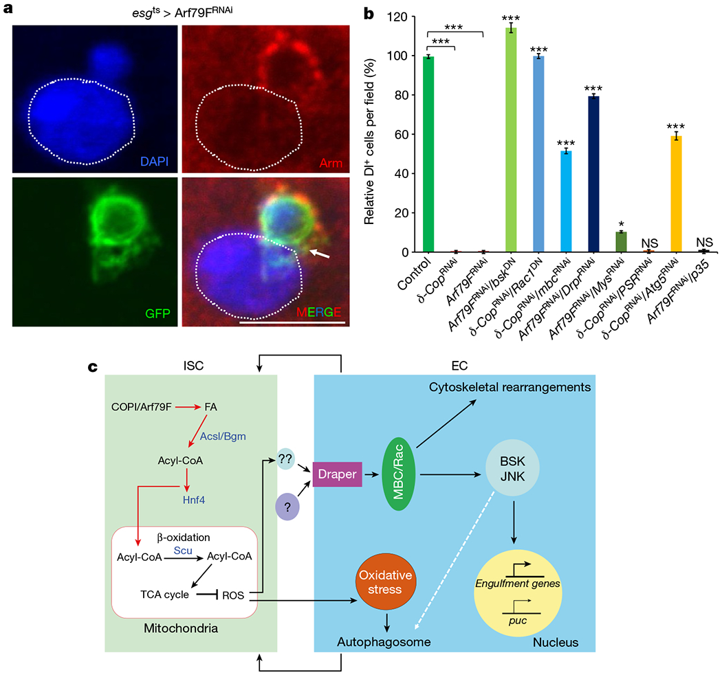
a, Representative images from esgts > Arf79FRNAi cells, 29°C, 7 d (n = 32). A dying ISC is engulfed by a neighbouring enterocyte. The posterior midguts of flies were dissected, stained with the GFP and Armadillo (Arm) antibodies, and analysed by confocal microscopy. Arrow points to GFP+ stem cell/progenitors and the dotted line outlines a nucleus and cell membrane of an enterocyte. Scale bar, 10 μ m. b, Quantification of Dl+ (Delta+) ISCs cells in flies with the NP1ts esgts driver and indicated genotypes (See images in Extended Data Fig. 9 for details). Data are represented as mean ± s.e.m. Statistical significance determined by Student’s t-test, *P < 0.05, ***P < 0.0001. NS, not significant (P > 0.05). c, Model of ISC death induced by knockdown of the COPI–Arf1 complex. Details are described in the text. The autophagosome is involved in the last step of phagocytosis (degradation of internalized cargo)27, and autophagy may both function downstream of and be regulated by the drpr–Mbc–Rac1–JNK pathway.