
Extended Data Figure 1 |. Stem cells are resistant to apoptosis.
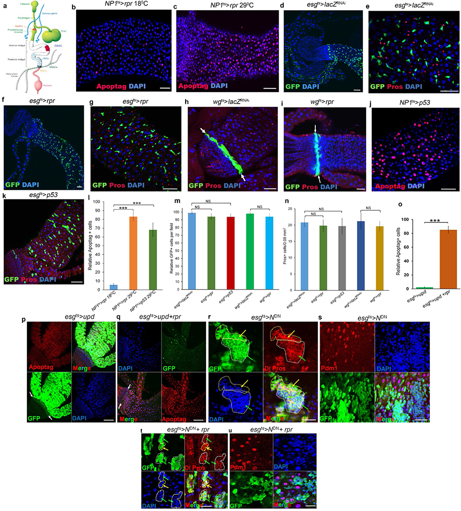
a, Stem cells in the adult Drosophila digestive system. In this system, three organs, the posterior midgut, the hindgut and the Malpighian tubules, meet and join at the junction of the posterior midgut and hindgut. Stem cells in these organs exhibit different degrees of quiescence. The intestinal stem cells (ISCs), located in the posterior midgut, divide once every 24 h2,3; the renal and nephric stem cells (RNSCs), located in the Malpighian tubules, divide about once a week4; and the quiescent hindgut intestinal stem cells (HISCs), found at the midgut–hindgut junction, divide only during stress-induced tissue repair5,6. GaSCs are gastric stem cells at the foregut–midgut junction47. GSSCs are gastric stem cells in the middle of the midgut48. The colours just make the cell types or organs more visible and do not exactly reflect different regions in the digestive system. b–n Stem cells are resistant to apoptosis. b, NP1ts > rpr, 18 °C, 24 h (n = 37). c, NP1ts > rpr, 29 °C, 24 h (n = 29). d, e, esgts > lacZRNAi, 29 °C, 7 d (n = 32). f, g, esgts > rpr, 29 °C, 7 d (n = 35). h, wgts > lacZRNAi, 29°C, 7 d (n = 27). i, wgts > rpr, 29°C, 7 d (n = 24). j, NP1ts > p53, 29 °C, 5 d (n = 31). k, esgts > p53, 29 °C, 7 d (n = 38). l, Quantification of Apoptag+ cells in the indicated panels. m, Quantification of GFP+ cells in the indicated panels. n, Quantification of Pros+ cells in the indicated panels. Data are represented as mean ± s.d.. Statistical significance determined by Student’s t-test, ***P < 0.0001. NS, not significant (P > 0.05). As reported previously8, 24-h induction of rpr in enterocytes resulted in widespread apoptosis (compare c with b and see the quantitative comparison in l). The induction of rpr by esg-Gal4 (f, g) or wg-Gal4 (i) had little effect on the progenitor or stem cells (that is, enteroblasts, ISCs, RNSCs and HISCs) at one week, compared to wild-type controls (compare f, g with d, e; i with h, and see the quantitative comparison in m). We also found that the overexpression of Drosophila p53 could effectively ablate the enterocytes in five days (compare j with b and see the quantitative comparison in l) but had little effect on stem cells even after one week, compared to controls (compare k with e and see the quantitative comparison in m). Because NP1–Gal4 and esg–Gal4 are not expressed in enteroendocrine cells, as expected, we did not find significant changes in enteroendocrine cells in these experiments (n). o–u, Activation of proliferation accelerates apoptotic cell death of hyperplastic stem cells but fails to completely eliminate neoplastic stem cells. o, Quantification of Apoptag+ cells in the indicated panels. Data are represented as mean ± s.d. Statistical significance was determined by Student’s t-test, ***P < 0.0001. p, esgts > upd, 29 °C, 4 d (n = 28). q, esgts > upd + rpr, 29°C, 4 d (n = 33). r, s, esgts > NDN, 29 °C, 7 d (n = 25). t, u, esgts > NDN + rpr, 29 °C, 7 d (n = 32). White arrows point to the hindgut–midgut junction in h, i, p, q; yellow arrows point to Pros+ enteroendocrine cells in r and t; green arrows point to Dl+ ISCs in r and t. White dotted lines outline GFP+ stem cell clusters in r and t. Yellow dotted lines outline enteroendocrine cell clusters in r and t. Expression of rpr or Arf79FRNAi in ISCs did not kill differentiated cells. The posterior midguts of flies with the indicated genotypes were dissected, stained with the indicated antibodies and analysed by confocal microscopy. Scale bars in b–k and p–u, 10 μm.