Fig. 1.
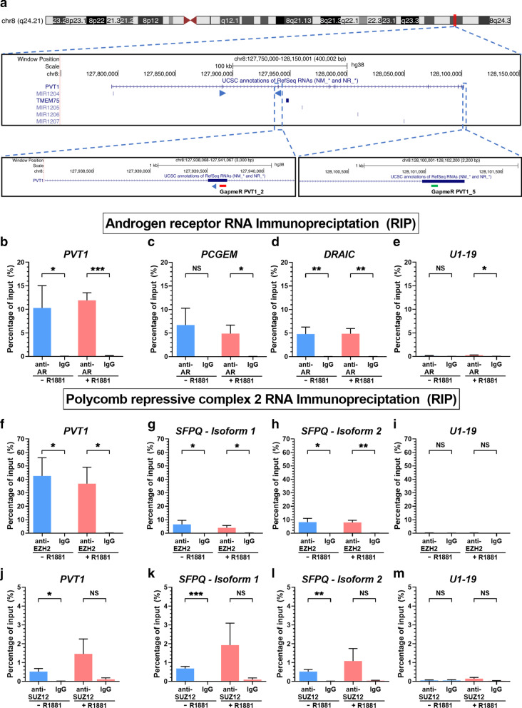
LincRNA PVT1 associated both with AR and EZH2 in hormone-starved or in androgen-stimulated LNCaP cells. a Snapshot of the PVT1 genomic locus on human Chromosome 8 showing the PVT1 lincRNA, along with the pair of PCR primers (blue arrowheads) that was used for its quantification. The two lower insets show the locations of two antisense LNA GapmeR oligos (red and green blocks) that were used for PVT1 knockdown in the experiments of Fig. 2. b–e RIP with anti-AR or nonspecific antibody (IgG), followed by RT-qPCR for genes PVT1 (b, target), PCGEM (c, positive control), DRAIC (d), and U1-19 (e, negative control). f–i, RIP with anti-EZH2 or nonspecific antibody (IgG), followed by RT-qPCR for PVT1 (f, target), for two different isoforms of SFPQ (g, h, positive controls) and U1-19 (i, negative control). j–m RIP with anti-SUZ12 or nonspecific antibody (IgG), followed by RT-qPCR for PVT1 (j, target), for two different isoforms of SFPQ (k, l, positive controls) and U1-19 (m, negative control). LNCaP cells under hormone-starved (-R1881, blue) or androgen-stimulated conditions (+ R1881, red) were tested. Data shown are mean ± s.e.m. of four biological replicates. T-test, *P < 0.05, **P < 0.01, ***P < 0.005

