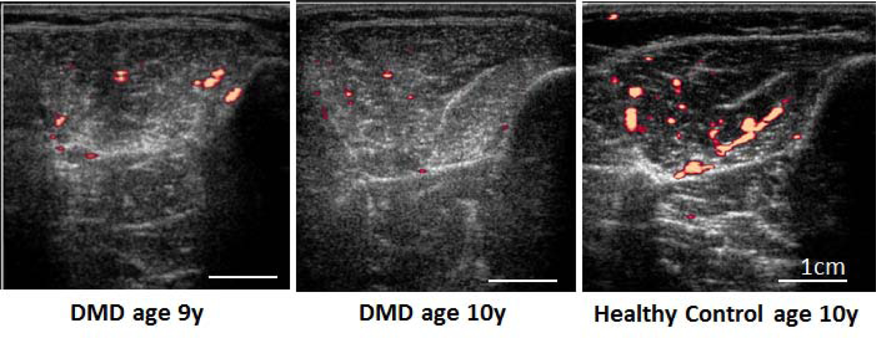
Figure 2: Post exercise blood flow in the Tibialis Anterior.

Power Doppler images of the tibialis anterior show reduced blood flow after exercise in a 9 and 10 year old boy with DMD compared to a healthy 10 year old boy.

Power Doppler images of the tibialis anterior show reduced blood flow after exercise in a 9 and 10 year old boy with DMD compared to a healthy 10 year old boy.