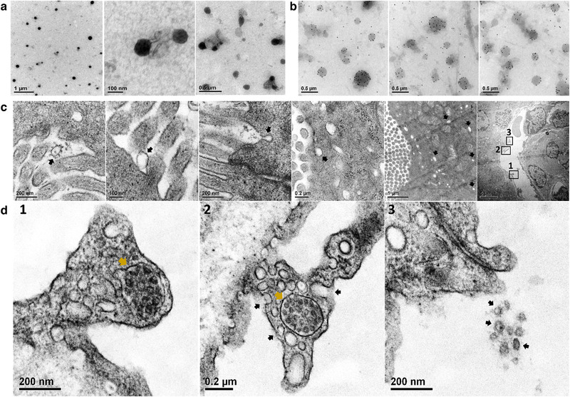
FIGURE 1.

Transmission electron microscopy (TEM) of casein micelles with CDC‐EV (a) immuno‐gold labelled CDC‐EV (b) and mdx mouse duodenum ∼10 min after oral gavage delivery of immuno‐gold labelled CDC‐EV with casein (c and d). Immuno‐gold labelled CDC‐EV were pointed with black arrows in the duodenal lumen, at and inside duodenal epithelia cell and inside duodenal vessel (c). D1‐3: Enlarged TEM images of a duodenal vessel (row C last image) showing immuno‐gold labelled CDC‐EV in a multi vesicular body next to vessel wall (MVB, d1, d2 gold arrows), immuno‐gold labelled CDC‐EV fusing with vessel wall (d2, black arrows) and, intravascular immuno‐gold labelled CDC‐EV (d3, black arrows)
