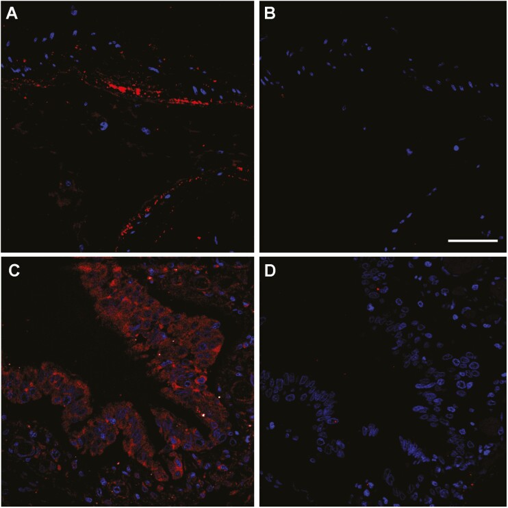
Figure 2.
Representative confocal microscopy images of lung tissue sections from non-COVID-19 autopsy specimens. Figure shows RAMP1 immunoreactivity (red) in the smooth muscle cells of an artery (A and B) and in the bronchiolar epithelium (C and D). Absence of the primary antibody (B and D) was used as a negative control. Cell nuclei were counterstained with DAPI (blue). Scale bar = 50 µm.

