
Figure 2. The commensal microbiota regulates IFNβ expression by dendritic cells in the colon LP.
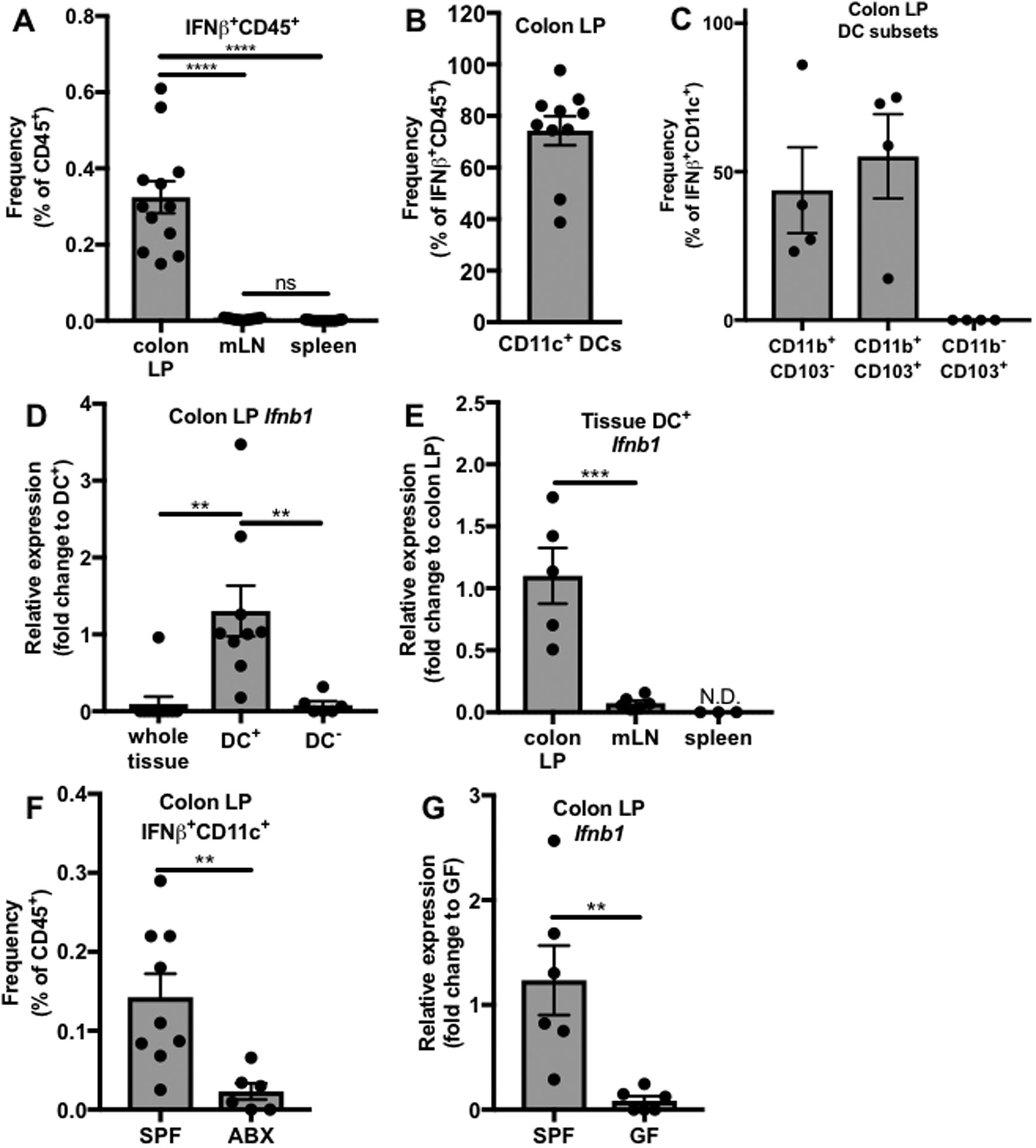
(A-C) Single cell suspensions were prepared from spleens, mLNs, and colon LP of SPF IFNβ-YFP reporter mice and analyzed by flow cytometry. (A) Frequency of IFNβ-YFP+ cells out of CD45+ cells (colon LP N=12, mLN N=13, spleen N=13). (B) Frequency of CD11c+ DCs out of total IFNβ-YFP+CD45+ cells in the colon LP (N=10). (C) Frequency of CD11b+CD103−, CD11b+CD103+, and CD11b−CD103+ DC subsets out of IFNβ-YFP+CD11c+ colonic LP dendritic cells. (D-E) DCs were isolated from single cell suspensions of different tissues from WT GF and WT SPF mice, yielding DC+ and DC− fractions, and Ifnb1 expression was analyzed by qRT-PCR. Relative expression of Ifnb1 in (D) the whole tissue (N=10), DC+ (N=9), and DC− (N=6) fractions of the colon LP or (E) the DC+ fraction of the colon LP (N=5), mLN (N=6), or spleen (N=3). (F) Flow cytometric analysis of frequency of colon LP IFNβ-YFP+CD11c+ cells out of CD45+ cells (ABX N=7, SPF N=10) in SPF or ABX treated IFNβ-YFP reporter mice. (G) qRT-PCR analysis of Ifnb1 expression in colon LP DC+ cells from WT SPF (N=6) or WT GF mice (N=6). (D-E,G) Fold change gene expression was calculated using the ΔΔCT method, with Actb as the reference gene, compared to the (D) DC+, (E) colon LP, or (G) SPF samples. Bars represent mean +/− SEM. Statistical analysis with (A,D,E) one-way ANOVA followed by Tukey’s multiple comparisons test and (F,G) unpaired t-test. N= number of mice, N.D.=not detected, *p<0.05, **p<0.01, ***p<0.001, ****p<0.0001, ns=not significant.