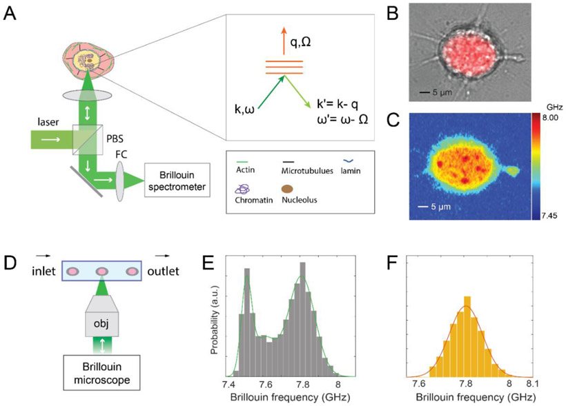
Figure 1. Characterization of nuclear mechanics with Brillouin technique.
(A) Principle of using a confocal Brillouin microscope to measure nuclear mechanics of a cell. The light red and yellow regions indicate cytoplasm and nucleus, respectively. (inset) the phase-matching condition indicates that the induced Brillouin frequency shift is equal to the frequency (a few GHz) of hypersonic acoustic waves Ω. ω and k represent the frequency and wave vector of incident light, and ω' and k' are the frequency and wave vector of scattered light, respectively. (B) co-registered bright-field and florescent image of a cell with its stained nucleus. (C) corresponding Brillouin image of the same cell. The color bar has a unit of GHz. (D) schematic of Brillouin flow cytometry. (E) original flow data of cell population with the fitting of the histogram’s profile. (F) extracted signature of the nucleus. The details of the data extraction procedure are provided in Methods.

