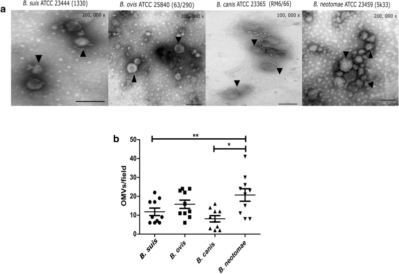
Fig. 2.
Purified OMVs from B. suis, B. ovis, B. canis and B. neotomae observed by electronic microscopy. a OMVs stained with phosphotungstic acid showed vesicles with a lipid bilayer membrane (arrowheads). b Graph representing the number of vesicles counted from ten fields for each strain (one-way ANOVA, 95% confidence interval). A significant difference was observed. *P < 0.05, **P < 0.01. Bar = 100 nm

