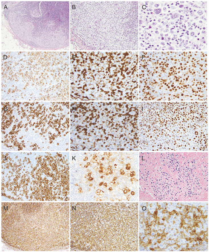
Figure 2.
Morphologic and immunophenotypic features of the lymphoma involving left axillary lymph node. A-C. H&E morphology (A, x40; B, x100; C, x400). D-K. Immunohistochemical stains (D, CD20; E, CD3; F, PAX-5; G, CD23; H, CD5; I, LEF1; J, CD30; K, CD15). L. EBER in situ hybridization. M-O. Immune check point antigen expression (M, PDL-1; N, HLA-1; O, B2M).

