. 2021 Jan 4;10:e60175. doi: 10.7554/eLife.60175

Figure 4. SUN1 KASH-lid residues involved in 6:6 assembly are essential for KASH1-binding.

(a,b) Gel filtration analysis. GCN4-SUN1 and MBP-KASH proteins were co-expressed and purified by amylose affinity (utilising non-specific binding by SUN1 for non-interacting mutants) and ion exchange (Figure 4—figure supplement 1d), and all fractions containing SUN-KASH complexes and dissociated proteins were concentrated and loaded onto an analytical gel filtration column. The elution profiles were validating by SEC-MALS in which wild-type fusion complexes and dissociated GCN4-SUN1 and MBP-KASH1 proteins were found to be 6:6 complexes, trimers and monomers, respectively (Figure 4—figure supplement 1f). (a) Gel filtration chromatograms (UV absorbance at 280 nm) across elution profiles for SUN1 wild-type (WT; dark blue), I673E (red), F671E (light blue), and W676E (green), with KASH4 (left), KASH5 (middle), and KASH1 (right), and (b) SDS-PAGE of their corresponding elution fractions. Representative of three replicates using different protein preparations. Source data are provided in Figure 4—source data 1.

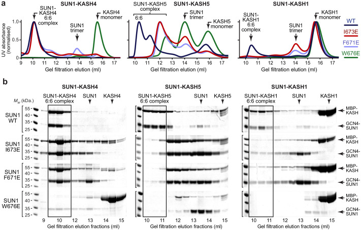
Figure 4—source data 1. Uncropped gel images relating to Figure 4b.

Figure 4.

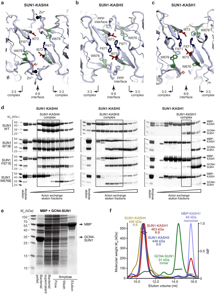
Figure 4—figure supplement 1. SUN-KASH complex formation upon SUN1 KASH-lid mutagenesis.

Figure 4—figure supplement 1.

(a–c) Structural details of the 6:6 interface of (a) SUN1-KASH4, (b) SUN1-KASH5, and (c) SUN1-KASH1, highlighting SUN1 KASH-lid residues I673 (red), F671 (blue), and W676 (green). (d) SDS-PAGE of anion exchange elution profiles of GCN4-SUN1 and MBP-KASH proteins that were co-expressed and purified by amylose affinity (utilising non-specific binding by SUN1 for non-interacting mutants), comprising SUN1 wild-type, I673E, F671E, and W676E, with KASH4 (left), KASH5 (middle), and KASH1 (right). All fractions containing SUN-KASH complexes or dissociated proteins were pooled, concentrated and loaded onto an analytical gel filtration column for the analyses shown in Figure 4a,b. (e) Amylose pulldown following co-expression of MBP and GCN4-SUN1, showing that GCN4-SUN1 binds non-specifically to amylose resin. (f) SEC-MALS analysis demonstrating that SUN1-KASH1/4/5 fusion complexes, GCN4-SUN1 and MBP-KASH1 are 6:6 complexes, trimers and monomers, respectively (theoretical masses – 466, 464, 464, 88, and 48 kDa). This analysis provides validation for the gel filtration elution profiles shown in Figure 4a,b.
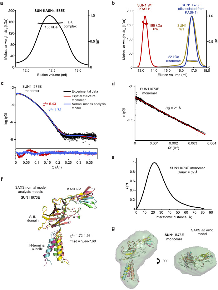
Figure 4—figure supplement 2. Biophysical analysis of the SUN1 I673E mutant.

Figure 4—figure supplement 2.

(a) SEC-MALS analysis demonstrating that SUN1 I673E and KASH4 form a 155 kDa 6:6 complex (theoretical 6:6 – 155 kDa). (b) SEC-MALS analysis demonstrating that isolated SUN1 wild-type (WT; yellow), and SUN1 I673E that has dissociated following co-expression with KASH1 (blue), are 22 kDa monomers (theoretical – 23 kDa). The wild-type SUN1-KASH1 6:6 complex is shown in red for comparison. (c–g) SAXS analysis of the SUN1 I673E monomer. (c) SAXS scattering curve overlaid with theoretical scattering curves of a SUN1 protomer from the SUN1-KASH1 crystal structure (red; χ2 = 5.43) and its normal mode analysis model (blue; χ2 = 1.72). (d) SAXS Guinier analysis revealing a radius of gyration (Rg) of 21 Å; the linear fit is shown in black and demarcated by dashed vertical lines (Q.Rg values were <1.3). (e) SAXS P(r) distribution showing a maximum dimension of 82 Å. (f) SAXS normal mode analysis in which the five highest scoring models are displayed, based on their fit to the experimental SAXS data (χ2 = 1.72–1.98), and demonstrate hinge-like motion of the N-terminal α-helix relative to the SUN domain. (g) SAXS ab initio model (NSD = 0.645; reference model χ2 = 1.85) into which the normal mode analysis models are docked.