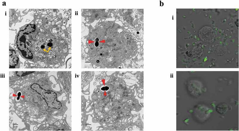
Figure 1.

Infection of macrophages by E. faecalis and their intracellular localization within the cytoplasm. A. TEM images of E. faecalis infection prior to macrophage differentiation showing i. bacteria within the cytoplasm of a macrophage (yellow arrows), ii–iv. Sections with bacterial replication (binary fission; red arrows) within intracellular vacuoles. Bi-ii. immunofluorescent images with the uptake of CFSE-stained E. faecalis by fully differentiated macrophages
