Fig. 2.
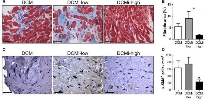
Increased PAI-1 level in DCMi-high patients attenuates fibrosis by inhibiting the activation of cardiac myofibroblasts. a Representative photomicrographs from cardiac sections of patients with DCM, DCMi low-grade inflammation (DCMi-low) and DCMi high-grade inflammation (DCMi-high) stained with Trichrome stain AB solution. Fibrous connective tissue is identified in blue. Magnification: ×400. Scale bar: 20 µm. b Quantification of fibrotic area (n = 6–9 patients per group). c Representative photomicrographs from cardiac sections immunostained with anti α-smooth muscle actin (α-SMA) antibody. α-SMA+ myofibroblasts are identified in brown. Magnification: ×600. Scale bar: 40 µm. d Quantification of α-SMA+ cells (n = 6–12 patients per group). Data are represented as mean ± SEM. *p ≤ 0.05 vs. DCM and DCMi-low or as indicated

