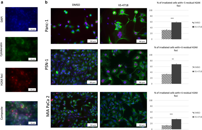
Fig. 4.
a Effect of VS-4718 on γ‑H2AX foci in Panc‑1 cell line in coculture with PSCs 24 h after radiation (VS-4718 2.5 µM was added 1 h before 6 Gy radiotherapy), cells were fixed labeled against γ‑H2AX, nuclei were stained with DAPI and PCCs were labeled against cytokeratin 8, then images were taken at 20 × objective, scale bar = 50 µm, finally a composite from three images (DAPI, cytokeratin‑8, and γ‑H2AX) was generated using ImageJ. b Composite images from three cell lines with bars represent the percentage of cancer cells with more than 5 or 6 foci per nucleus, scale bar = 50 µm. Data shown as mean ± standard deviation, n = 3; t-test; **P < 0.01, ***P < 0.001

