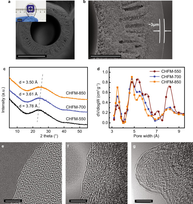
Fig. 2. Morphology and structure characterization of the carbon hollow fiber membranes (CHFMs).
a, b Cross-sectional SEM images of CHFM-700 (carbonized at 700 °C) (scale bars: a 100 μm, b 20 μm), and the inset shows a CHFM with a bend radius of <1.5 cm (scale bar: 1 cm), indicating mechanical flexibility. The thickness of a selective layer is ca. 3 μm, and the porous morphology is well-maintained after carbonization; c the XRD patterns of CHFMs carbonized at different temperatures. The d-space was calculated from the Bragg equation, and found to decrease from 3.78 to 3.5 Å with the increase of final carbonization temperature from 550 to 850 °C, and d the pore size distributions of CHFMs calculated by the NLDFT model from CO2 physisorption at 0 °C. The specific volumes of ultramicropores (especially the pores of <4 Å) for CHFM-850 and CHFM-700 are larger compared to that of CHFM-550, indicating more diffusion (size)-selective membranes; e–g HR-TEM images of CHFM-550, CHFM-700, and CHFM-850, respectively. Scale bar: 5 nm.

