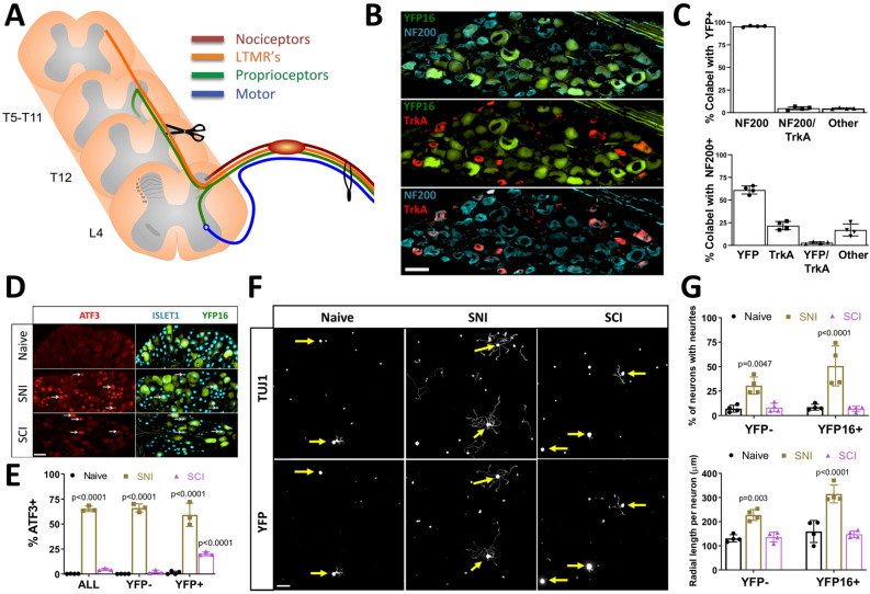
Figure 2.
Spinal Cord Injury (SCI) does not induce a pro-growth state in ascending DRG sensory neurons in vitro. (A) Schematic of the experimental design and peripheral and central projects of major sensory neuron subtypes of L4 dorsal root ganglion (DRG) neurons. (B) Representative images of L4 dorsal root ganglion (DRG) neurons from Thy1YFP16 mice labeled with NF200 (blue) and TrkA (red) antibodies. (C) Quantification of (B) indicating percent overlap of YFP, NF200, and TrkA labeling in Thy1YFP16 mice (n = 4 mice). (D) Representative images of L4 DRG neurons labeled with ATF3 and Islet1 antibodies in Thy1YFP16 mice in naive or 3 days after sciatic nerve injury (SNI) or SCI. (E) Quantification of (D) indicating percentage of ATF3 positive, Islet-1 labeled neuronal nuclei in all neurons, as well as YFP negative and YFP positive neurons, for each condition (n = 3 mice/group; 2-way ANOVA). White arrowheads point to ATF3 positive neuronal nuclei. (F) Representative images of cultured cells from L4 DRG labeled for all neurons (TUJ1) and YFP positive neurons in naïve or 3 days after SNI or SCI. (G) Quantification of (F) indicating the percentage of neurons extending neurites and the average radial length of neurites, in YFP negative and YFP positive neurons, for each condition (n = 4 mice/group; 2-way ANOVA). Yellow arrows point to the cell body of YFP positive neurons.

