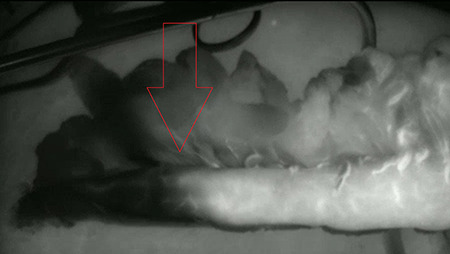
Fig. 1.

Gastric conduit before transfer to cervical region. This is a near infrared fluorescence view with the fluorescent signal displayed in white. A clear demarcation is noticed at the red arrow. The anastomosis was constructed within the fluorescent area.
