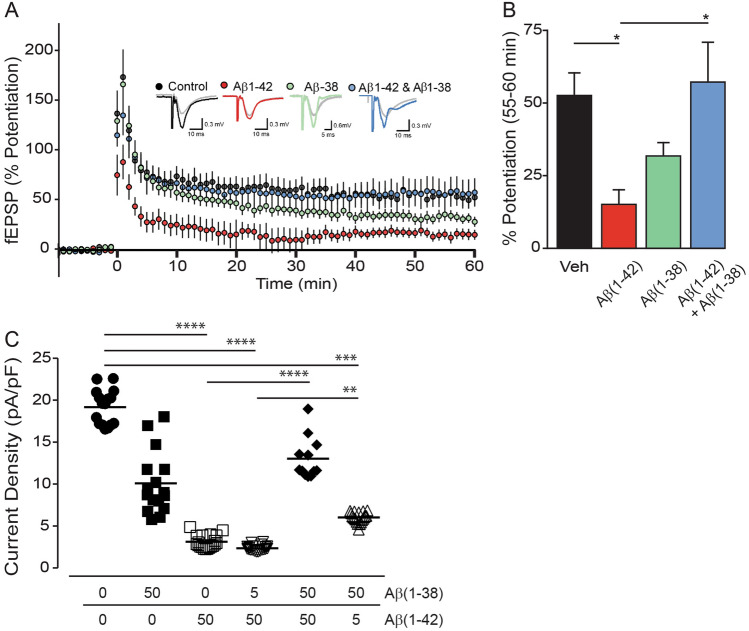
Figure 5.
Aβ(1–38) rescues the deficit caused by application of Aβ(1–42) in electrophysiological paradigms. (A) Long-term potentiation (LTP) graph showing the percent potentiation (mean ± sem) following theta burst LTP in control (black; n = 10), Aβ(1–42) (red; n = 8), Aβ(1–38) (green; n = 6), and Aβ(1–42) + Aβ(1–38) (blue; n = 7) conditions (all peptides at 500 nM). Theta burst stimulation was applied at time = 0 min. Representative fEPSP responses obtained during the baseline (grey traces) and 55–60 min following LTP induction (black, red, blue and green traces) are shown for each condition. (B) Bar graph showing the percent potentiation (mean ± sem) 55–60 min post-LTP induction. *: P < 0.05. (C) Average current densities (± sem) from whole cell voltage-clamp recordings in primary hippocampal neurons exposed to Aβ(1–38) and Aβ(1–42) (in µM, 24 h). Cells were held at -60 mV and currents were elicited by voltage ramps, e.g., step depolarized between -80 and + 100 mV (in 20 mV increments). Current density was measured at 0 and 20 mV, 100 ms after the voltage step. Currents were divided by cell capacitance and reported as current densities (pA/pF). **: P < 0.01; ***: P < 0.001; ****: P < 0.0001 between indicated groups.

