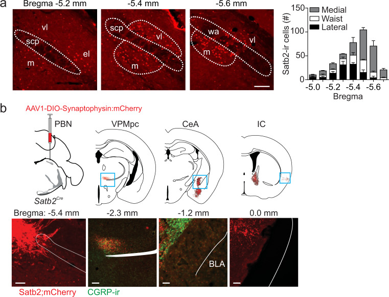
Fig. 1. Satb2 neurons are expressed in the gustatory PBN.
a Immunostaining for Satb2 in the PBN and quantification of its expression across the rostral–caudal extent of the PBN (n = 3 mice). scp superior cerebellar peduncle, vl ventral–lateral, m medial, wa waist. b Unilateral injection of AAV1-DIO-synaptophysin:mCherry in the PBN of Satb2Cre mice and labeling of terminal fields in select brain regions (n = 3 mice). The Satb2–neuron projection overlaps with CGRP expression in the VPMpc, but not in the CeA. Scale bars, 100 µm. Data are presented as mean ± SEM. Source data are provided as a Source Data file.

