Figure 2.
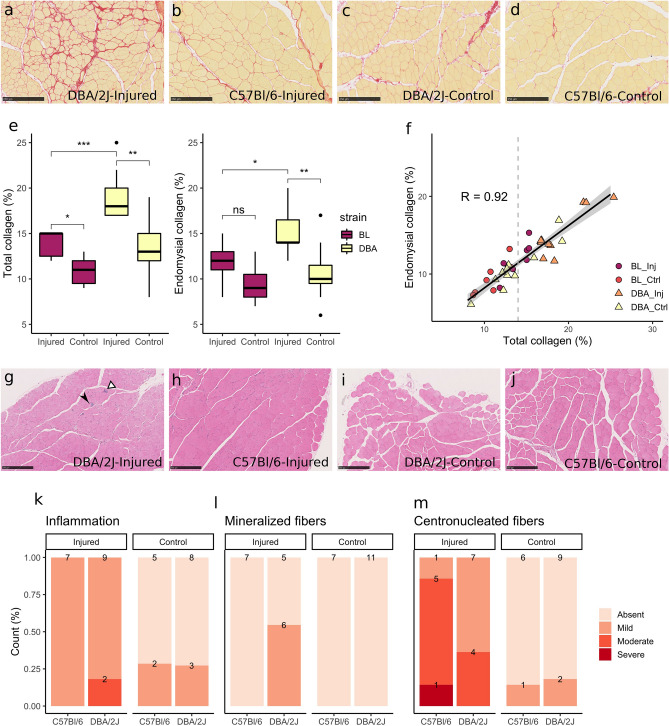
Injury induced fibrosis in non-dystrophic muscles. (a–d) Sirius red collagen staining of (a) DBA/2J injured muscle, showing marked fibrosis in red; (b) C57BL/6 injured muscle, with a moderate increase in collagen staining; (c) DBA/2J and (d) C57BL/6 non-injured control muscles, showing thin connective tissue layers characteristics of normal skeletal muscles. (e) Boxplots show increased total and endomysial collagen fraction in injured muscles, especially in DBA/2J mice. (f) Total and endomysial collagen fractions were highly correlated (R = 0.92). The majority of non-injured samples presented total collagen fraction below 14%, while the injured muscles presented collagen fraction higher than 14% in general. (g–j) Haematoxylin–eosin staining of (g) DBA/2J injured muscle, with moderate inflammation (white arrowhead) and presence of mineralized fibers (black arrowhead); (h) C57BL/6 injured muscle, showing high count of centronucleated fibers but no inflammation; (i) DBA/2J and (j) C57BL/6 non-injured control muscles. (k–m) Increased collagen fraction occurred simultaneously with mild inflammation (k), low count of necrotic mineralized fibers in DBA/2J mice (l) and regeneration, reflected by the presence of centronucleated fibers (m). (a), (c): magnification × 10, scale bar 250 μm. *p < 0.05, **p < 0.01, ***p < 0.001, ns non-significant.

