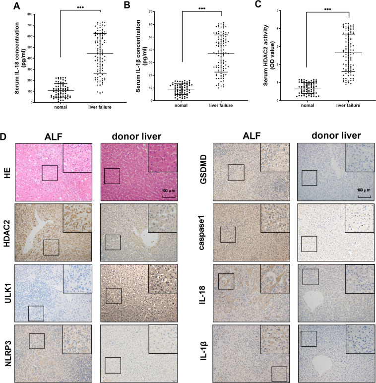
Fig. 6. Serum IL-18, IL-1β, HDAC2 level, pathological changes, and protein expression levels of HDAC2, NLRP3, GSDMD, caspase 1, IL-18, and IL-1β in liver tissues of normal people and ALF patients.
A–C The serum IL-18, IL-1β, and HDAC2 level in normal people and ALF patients. The level of HDAC2, IL-18, and IL-1β in liver failure was higher than normal donors. D Normal liver tissue showed neatly arranged hepatocytes with no inflammatory cell infiltration. Hepatocytes in the liver failure tissue had a lot of necrosis and inflammatory cell infiltration, and the tissue also contained accumulated red blood cells. The expression levels of HDAC2, NLRP3, GSDMD, caspase 1, IL-18, and IL-1β in ALF patients’ liver tissues were higher than the normal controls. The expression of ULK1 protein expression was reduced. Transmission electron microscopy showed that the nucleus and cell membrane structure were incomplete in the liver failure tissues, and the mitochondria were swollen with a small amount of autophagosomes. Data are expressed as the mean ± SEM. N = 70 in normal group, and n = 106 in ALF group. ***P < 0.01.

