Figure 10.
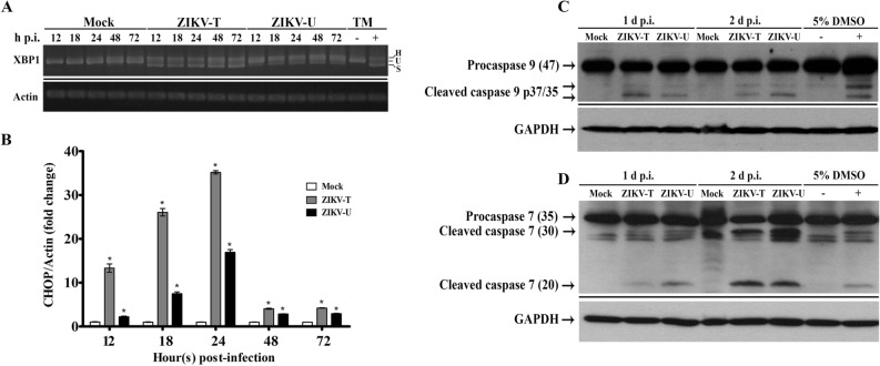
Activation of the UPR and induction of apoptosis during ZIKV infection. A549 cells were mock-infected or infected with ZIKV-T or ZIKV-U at MOI 5 or 2, respectively. At various times as indicated total RNA was extracted and examined for (A) the presence of the unspliced (U), spliced (S) and heteroduplex (h) forms of XBP1 mRNA using RT-PCR, with tunicamycin (TM, 4 μg/ml) treated cells as a splicing control and actin as internal control. The PCR products were separated by electrophoresis on 2.0% agarose gels and different PCR experiments are separated by a continuous black line, or (B) expression of CHOP mRNA using quantitative real time PCR. The relative expression levels of CHOP mRNA were normalized against actin using the comparative CT method (2−ΔΔCT method). Error bars represent SEM. (*p value < 0.05). Total protein was collected and subjected to western blot analysis to determine the proteolytic processing of (C) caspase 9, and (D) caspase 7. GAPDH was used as an internal loading control. Cells treated or untreated with tunicamycin or DMSO were included as positive and negative controls. Different probings of the same filter are separated by a continuous black line and uncropped blots are presented in Supplemental materials.

