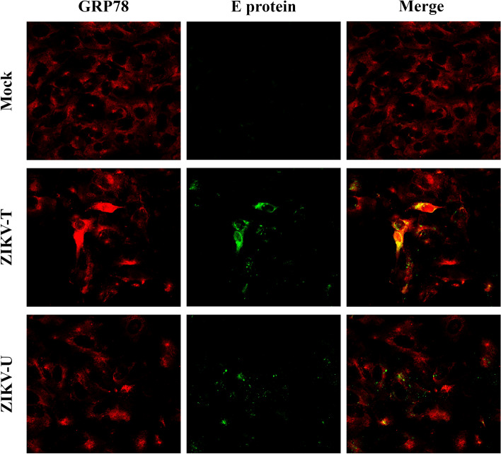
Figure 5.
Colocalization analysis of GRP78 and ZIKV E protein on the cell surface. A549 cells were grown on glass slides and then incubated with ZIKV-T or ZIKV-U at 4 °C for 1 h. Under non-permeabilization conditions, cells were strained with primary antibodies followed by appropriate secondary antibodies. The cell surface colocalization (yellow) between GRP78 (red) and ZIKV E protein (green) was determined using a ZEISS confocal microscope.

