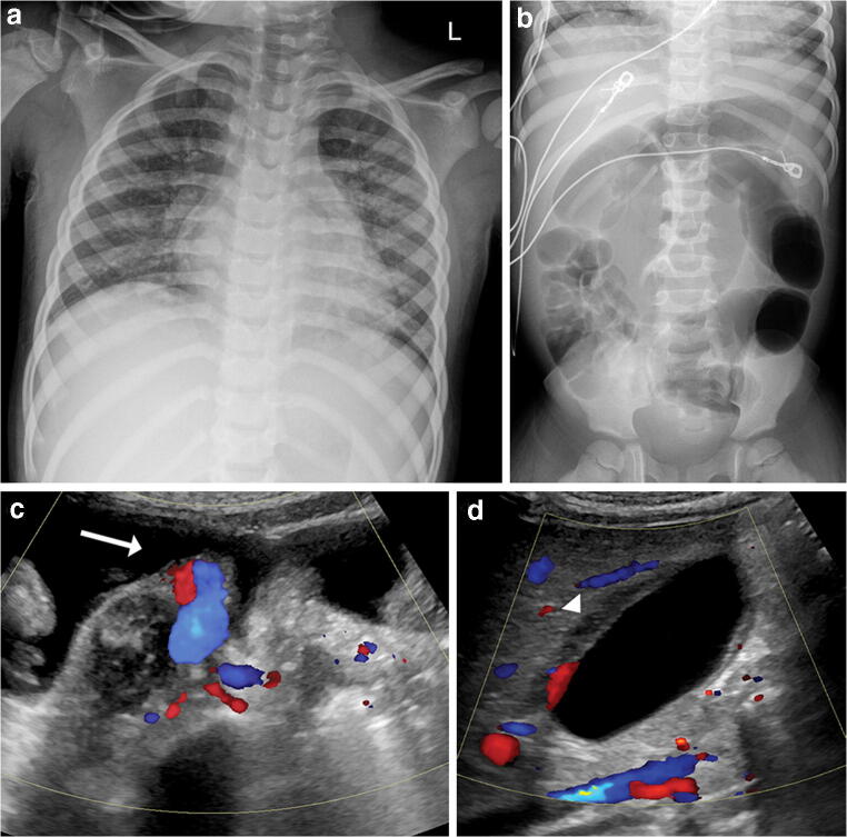
Fig. 1.
Imaging in a 4-year-old boy with a medical history of prematurity and mild asthma who presented with fever, abdominal pain, diarrhea, hypotensive shock and evidence of myocardial dysfunction with positive coronavirus disease 2019 (COVID-19) reverse transcription polymerase chain reaction (RT-PCR) and serology results. a Anteroposterior chest radiograph demonstrates bilateral perihilar opacities and peribronchial thickening. b Supine radiograph of the abdomen demonstrates abdominal distention with gaseous distention of the colon down to the rectum, suggesting colonic ileus. c Transverse US image of the right lower abdominal quadrant at the level of the iliac vasculature demonstrates small-volume ascites (arrow). d Sagittal sonographic image of the gallbladder demonstrates gallbladder wall thickening (arrowhead) but no gallstones

