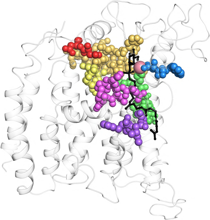
Fig. 4.
Sequence motifs M1-M7 come spatially together in model structures of TMTCs. We illustrate the spatial localization of sequence motifs M1 (red), M2 (orange), M3 (yellow), M4 (green), M5 (blue), M6 (violet) and M7 (pink, all shown in ball mode) at the background of the structural cartoon of the whole protein. DPM is presented as blackish sticks, the divalent metal ion is represented as reddish sphere. We show the case of TMTC1; the figures for the other TMTCs look very similar. To note, motif M2 in this figure is extended to the conserved region represented by SHKSYRPLCVTLTSFRLN in TMTC1 (88–103 in EL1)

