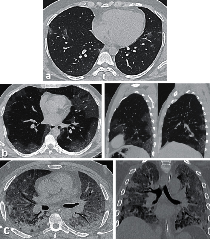
Figure 1.

(a) Axial thin sections of unenhanced CT chest scan show mild GGO involving bilateral peripheral lower lobes. (b) Axial and sagittal sections show bilateral peripheral multilobe GGO of moderate disease severity. (c) Axial and coronal sections show diffuse crazy-paving pattern with areas of peripheral consolidations indicating severe disease.
