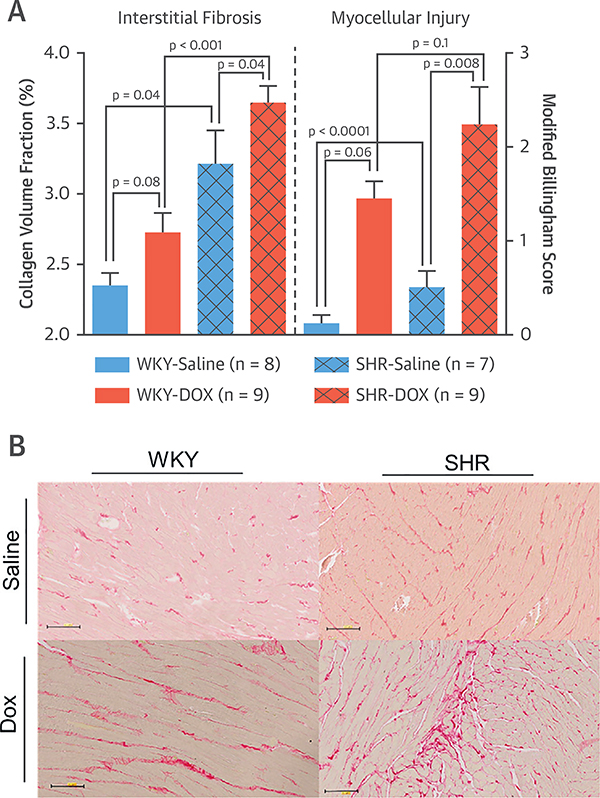
FIGURE 1. Histopathological Assessment of Cardiac Fibrosis and Myocellular Injury.
(A) Collagen volume fraction (CVF) (left y-axis) and modified Billingham Score for myocardial injury (right y-axis) of WKY rats treated with saline (solid blue bars) or DOX (solid orange bars) and SHRs treated with saline (hash-marked blue bars) or DOX (hash-marked orange bars). All values are mean ± SEM. (B) Representative microphotographs (20×) of left ventricles from each treatment group stained with Picrosirius red that identifies collagen. DOX = doxorubicin; SHR= spontaneously hypertensive; WKY = Wistar Kyoto.

