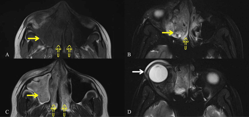
Figure 1. Orbital Cuts of MRI Brain.
almost complete opacification involving the right maxillary (solid yellow arrows) and bilateral ethmoid (yellow dotted arrows) sinuses, appearing (A) hypointense on T1 and (B, C) hyperintense on T2-weighted images. (D) There is mass effect causing slight proptosis of the right eyeball with preorbital soft tissue swelling (solid white arrow).
MRI: magnetic resonance imaging

