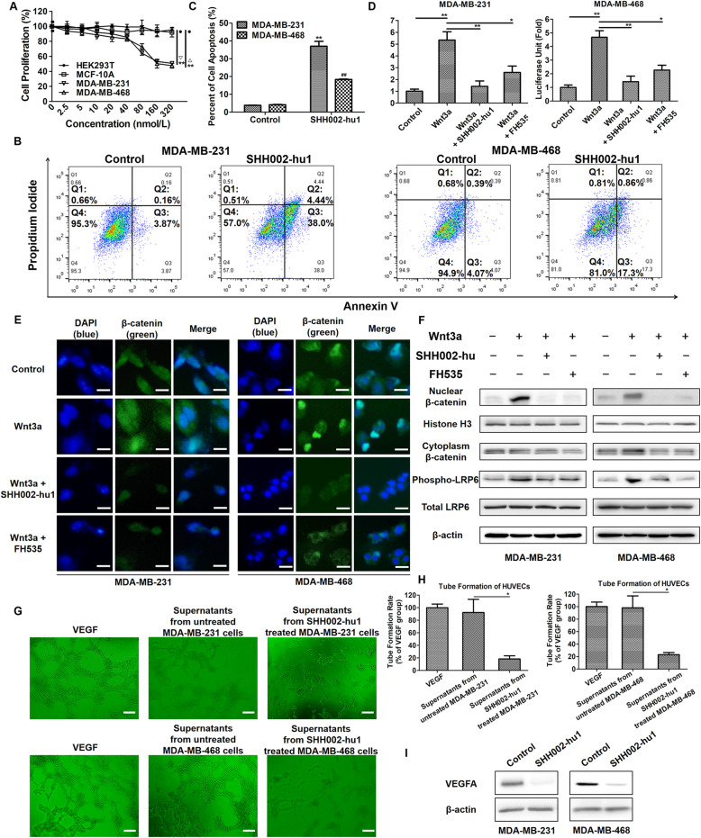
Fig. 3.
SHH002-hu1 inhibits the proliferation of TNBC cell lines and tumor angiogenesis through blocking Wnt/β-catenin pathway. a. The viability of MDA-MB-231/MDA-MB-468 cells was assessed by MTT assay at 48 h after treatment with different concentrations of SHH002-hu1. HEK293T cell and MCF-10A were set as a negative control. SHH002-hu1 specifically inhibited the growth of Fzd7+ TNBC cells induced by Wnt3a in a dose-dependent manner. Data were given as the mean ± SD (n = 3), **p < 0.01. b. Representative plots showing the apoptosis patterns of SHH002-hu1 treated MDA-MB-231/MDA-MB-468 cells, the percentage of cells in each quadrant was indicated. c. Quantitative analysis of apoptosis assay. Data were presented as the mean ± SD, n = 3, **p < 0.01 (MDA-MB-231 cells), vs. the previous group; ##p < 0.01 (MDA-MB-468 cells), vs. the previous group. d. TOP/FOP ratio in MDA-MB-231/MDA-MB-468 cells (stimulated by Wnt3a) treated with SHH002-hu1 for 24 h. FH535 was set as a positive control. The results from 3 independent experiments are expressed as mean ± SD of fold change, *p < 0.05, **p < 0.01. SHH002-hu1 effectively inhibited the β-catenin/TCF-4 transcriptional activity induced by Wnt3a. e. SHH002-hu1 repressed the accumulation of β-catenin in the nucleus induced by Wnt3a. IF stainings of β-catenin (green) were shown, and nuclei were counterstained with DAPI (blue), bar = 20 μm. f. SHH002-hu1 blocked nuclear accumulation of β-catenin and phosphorylation of LRP6. MDA-MB-231/MDA-MB-468 cells were treated with Wnt3a and SHH002-hu1, then western blot assay was conducted as indicated. Histone H3 was used as loading control for nuclear proteins, and β-actin was for cytoplasmic proteins. SHH002-hu1 attenuated the Wnt3a induced accumulation of β-catenin and inhibited the induction of phosphorylated (Ser1490) LRP6 by Wnt3a. g, h. HUVECs tube-like photomicrographs and quantitative analysis revealed that the angiogenesis in in vitro model was significantly inhibited by the 8 h-incubation of SHH002-hu1-treated TNBC cells supernatants, bar = 100 μm. The quantitative analysis of HUVECs tube formation (a complete polygon was considered as a tube) was based on the mean of the 5 regions of each group. Data were presented as the mean ± SD, n = 5, *p < 0.05. i. Western blot assay indicated that SHH002-hu1 reduced the expression of VEGFA in TNBC cells remarkably

