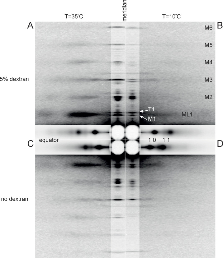
Figure 1.
Low-angle x-ray diffraction patterns from demembranated fibers from rabbit psoas muscle in relaxing conditions. (A–D) Patterns at 35°C (A and C) and at 10°C (B and D), in the presence (A and B) or absence (C and D) of 5% Dextran T500. The meridional and equatorial axes (parallel and perpendicular to the fiber axis) were digitally attenuated 2 and 20 times, respectively. Data added from three fiber bundles; total exposure time, 60 ms; specimen-to-detector distance, 3 m. Mi, myosin-based reflections indexing on its ∼43-nm axial repeat; ML1, first myosin layer line; T1, troponin-based reflection with axial periodicity ∼38 nm.

