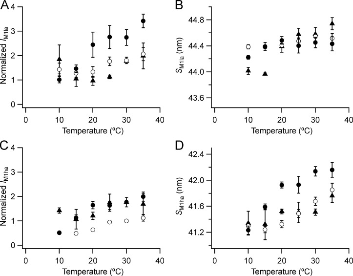
Figure S3.
Intensity and spacing of the component peaks of the M1 reflection. (A–D) Intensity (A and C) and spacing (B and D) are shown. la (A and B) and ha (C and D) denote lower- and higher-angle components. Mean ± SE. Open circles, no Dextran, n = 10 demembranated psoas fiber bundles; filled circles, 5% Dextran, n = 5; black triangles, resting intact mouse EDL muscles, n = 3. Intensities for demembranated fibers normalized by the value of IM1ha in the absence of Dextran at 30°C and those for intact muscles scaled to the value for demembranated fibers at 30°C in the presence of Dextran.

