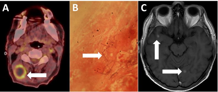
Figure 2.
(A) PET CT demonstrating high uptake in a right cervical lymph node (white arrow). (B) Gram stain of a lymph node aspirate demonstrating the beaded and branching Gram-positive rod, Nocardia farcinica (white arrow). (C) MRI head with contrast demonstrating the decreasing size of enhancing brain lesions (white arrows). PET, positron emission tomography.

