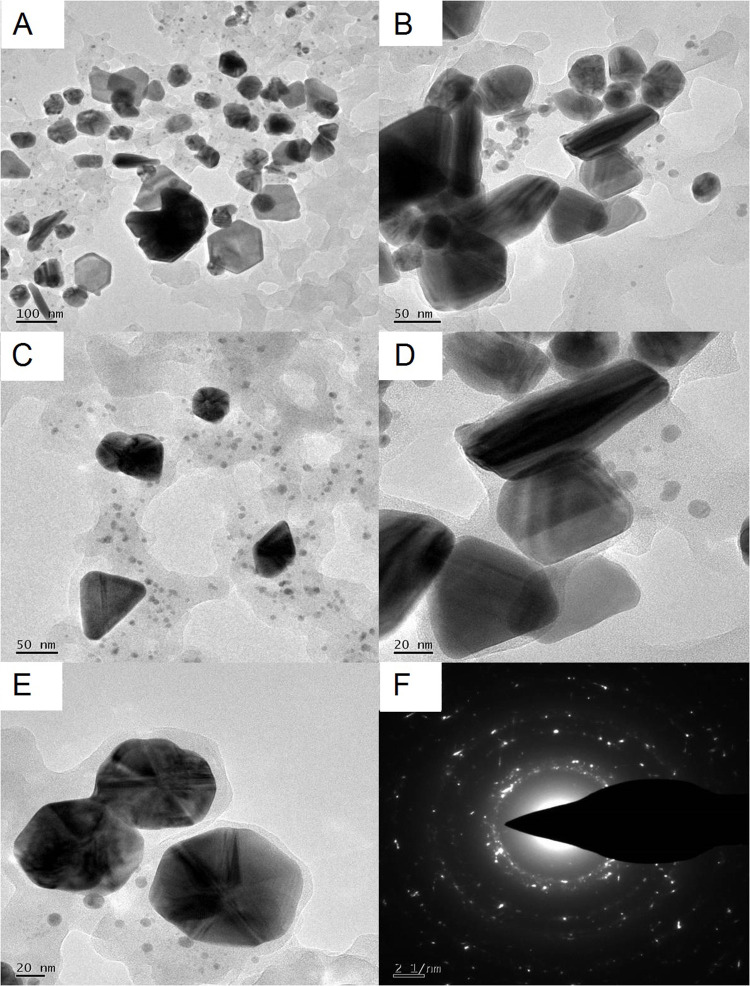
Figure 6.
TEM analysis PF@AgNPs (A) TEM micrograph at 100 nm shows monodispersed AgNPs with different shapes including spherical, rod, triangle, and rhombic; (B) rod-shaped, rhombic shaped, and spiral-shaped AgNPs observed at 50 nm scale; (C) triangle, rhombic and spherical shaped AgNPs at 50 nm; (D) rod-shaped and triangle-shaped AgNPs at 20 nm; (E) spherical shaped AgNPs at 20 nm; (F) SAED pattern of AgNPs.

