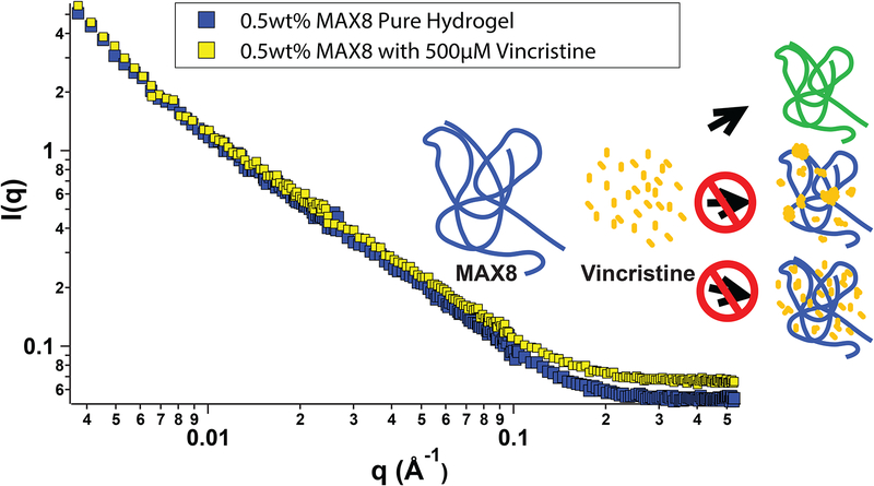
Figure 2 -.
Small-angle neutron scattering from 0.5 wt% MAX8 hydrogels with 500 μM vincristine (yellow) and without (blue) as a function of scattering variable q. Both lines have a similar overall shape and slope throughout the measured q range, implying that the presence of vincristine does not alter the structure of the MAX8 gel or the intramolecular folding of individual MAX8 chains. The cartoon inset shows the possible drug-gel configurations, a.) green fibrils indicating the yellow vincristine bound to the blue MAX8 fibrils, b.) domains of yellow vincristine mostly at the branch and entanglement points, or c.) yellow vincristine evenly scattered throughout the MAX8 network.

