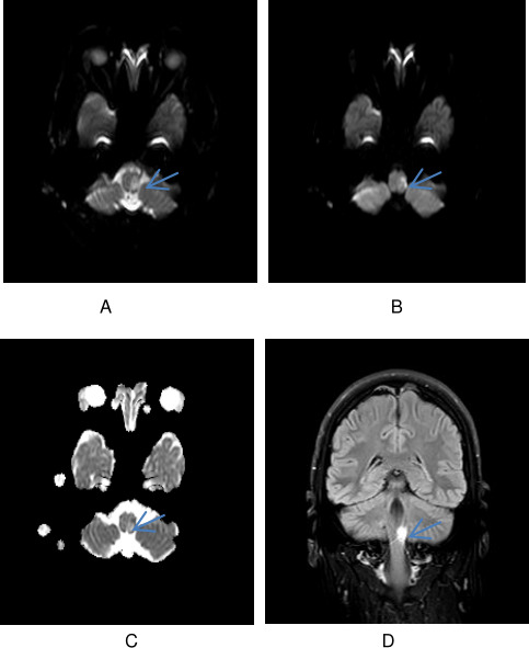
Figure 1.

(A) DWI b0 image of the lesion in the lateral part of the left medulla. (B) DWI b1000 image of the lesion in the lateral part of the left medulla. (C) ADC map demonstrating the variable diffusion restriction in the lesion suggestive of acute on a subacute infarct. (D) Coronal fluid attenuated inversion recovery (FLAIR) image showing an infarct in the left medulla.
