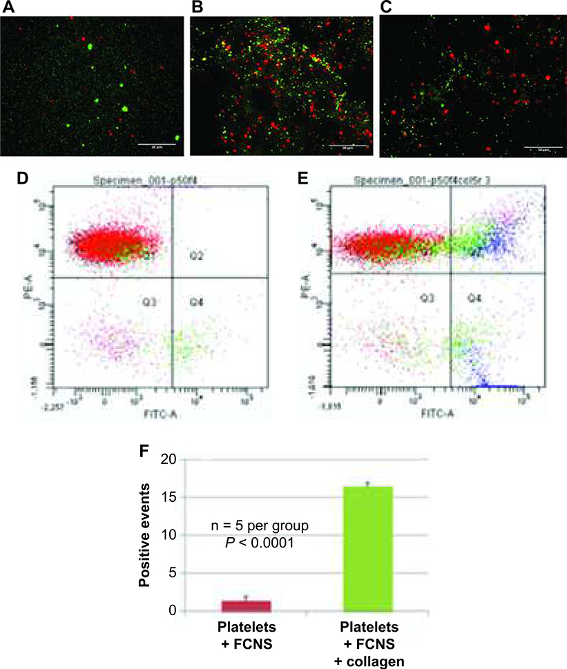
FIG. 3.
Fibrinogen-coated nanospheres (FCNs) bind to activated but not resting platelets. Fluorescence microscopy images of platelets (labeled red with CD9-PE antibody) and FCNs (labeled green with Alexa Fluor 488) show no interaction in the absence of platelet activation (panel A). The addition of collagen (a platelet agonist) shows yellow signal overlap (panel B), indicating interaction between the activated platelets and FCNs. However, control nanospheres (albumin spheres without fibrinogen coating) do not interact with platelets, even in the presence of collagen (panel C). The degree of interaction can be estimated with flow cytometry. When platelets (labeled CD9-PE) and FCNs (labeled Alexa Fluor 488) are mixed together in the absence of collagen, there are very few overlapping events, measured as double-positive events (panel D). When collagen is added, the number of double-positive events significantly increases (panel E). These data are quantified in panel F.

