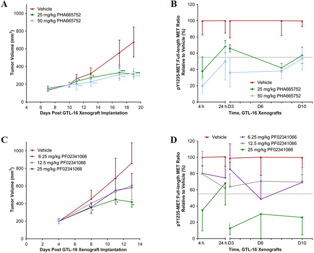
Figure 3. Reduction of GTL-16 tumor growth and MET phosphorylation by MET RTK inhibitors.
(A) Tumor volume of GTL-16 tumor xenografts in mice treated with PHA665752 at daily doses of 0, 25 and 50 mg/kg IP starting 10 days after implantation; n = 5-28 per dose per time point. (B) The intratumoral pY1235-MET:MET ratio during 10 days of daily treatment with PHA665752; n = 5-6 per dose per time point. Tumor samples were analyzed at 4 and 24 h after dose 1, and 4 h after dose 3 (D3), 8 (D8) and 10 (D10). (C) Tumor volume of GTL-16 xenografts in mice treated with PF02341066 at daily doses of 0, 6.25, 12.5, and 25 mg/kg PO starting at 4 days after implantation. Error bars are mean ± SD, groups n = 4-30 per dose per time point. (D) The pY1235-MET:MET ratio during 10 days of daily treatment with PF02341066. Tumor samples were analyzed at 4 and 24 h after dose 1 and 4 h after dose 3 (D3), 6 (D6) and 10 (D10). Mean ± SD, groups n = 2-6 per dose per time point. For (A) and (C), single asterisk (*) p < 0.05 and triple asterisk (***) p < 0.001 compared to vehicle group by unpaired Student’s t-test. For (B) and (D), the dotted line indicates the Least Significant Change (LSC) in pMET:MET ratio from the vehicle-treated group of 45% ; changes larger than this are attributed to drug effect.

