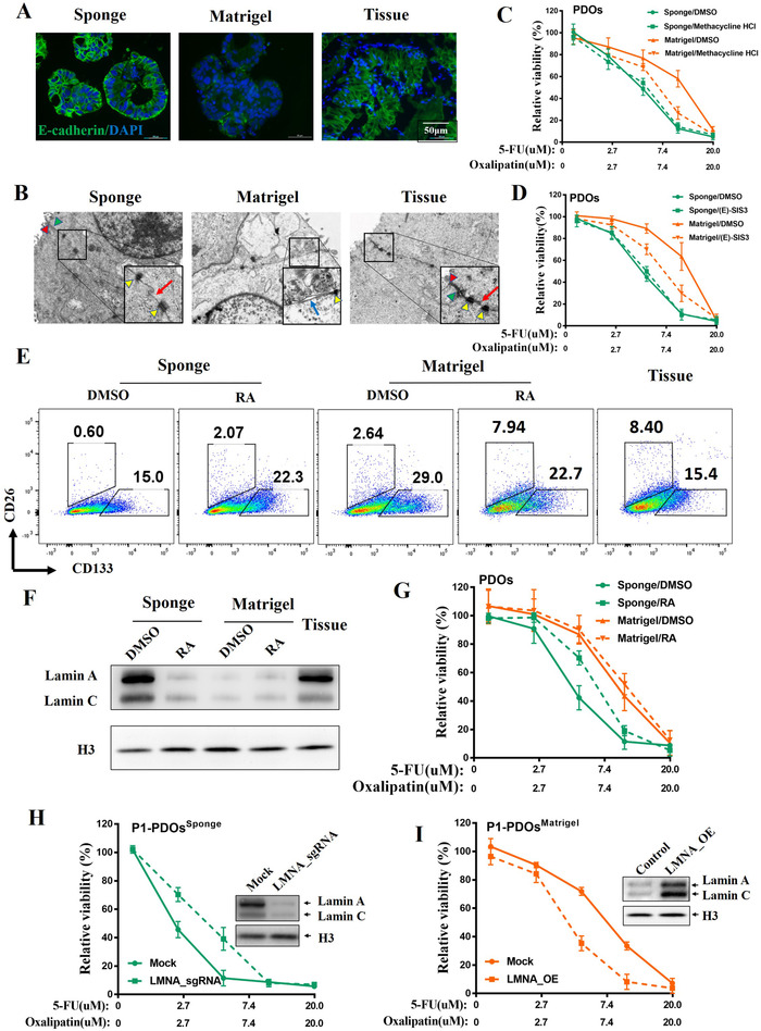
FIGURE 4.

HA‐Coll sponge can keep the epithelial and differentiation state of PDOs. (A) The expression levels of epithelial marker E‐cadherin of representative P1 were examined by immunofluorescence. The epithelial cells of parental biopsy were isolated as control. (B) Cell–cell junctions in PDOsSponge and PDOsMatrigel from representative P1 were examined by transmission electron microscopy. Red arrowhead for tight junctions; Green arrowhead for adherens junctions; yellow arrowhead for desmosome; red arrow for gap junctions; blue arrow for dissolution of cell–cell junctions. (C and D) The ex vivo dose‐response curves (DRCs) of representative PDOs from P1 exposed to FO regimen after adding the EMT inhibitors: Methacycline HCl (10 µM) or (E)‐SIS3 (3 µM). (E) Lamin‐A contribute to differentiation state of PDOsSponge. The proportion of CRC‐CSCs in PDOs and parental tissues from P1 was analyzed by flow cytometry. PDOs were pretreated with 5‐FU and oxaliplatin for 6 days with or without 1 µM retinoic acid (RA). (F) The expression levels of lamin A protein in PDOs and their parental tumor tissues from P1 were examined by immunoblotting. (G) The DRCs of PDOs from P1 treated with 5‐FU and oxaliplatin in the presence of RA. (H) The DRCs of PDOsSponge from P1 treated with 5‐FU and oxaliplatin after LMNA KO. (I) The DRCs of PDOsMatrigel from P1 treated with 5‐FU and oxaliplatin after LMNA overexpressed
