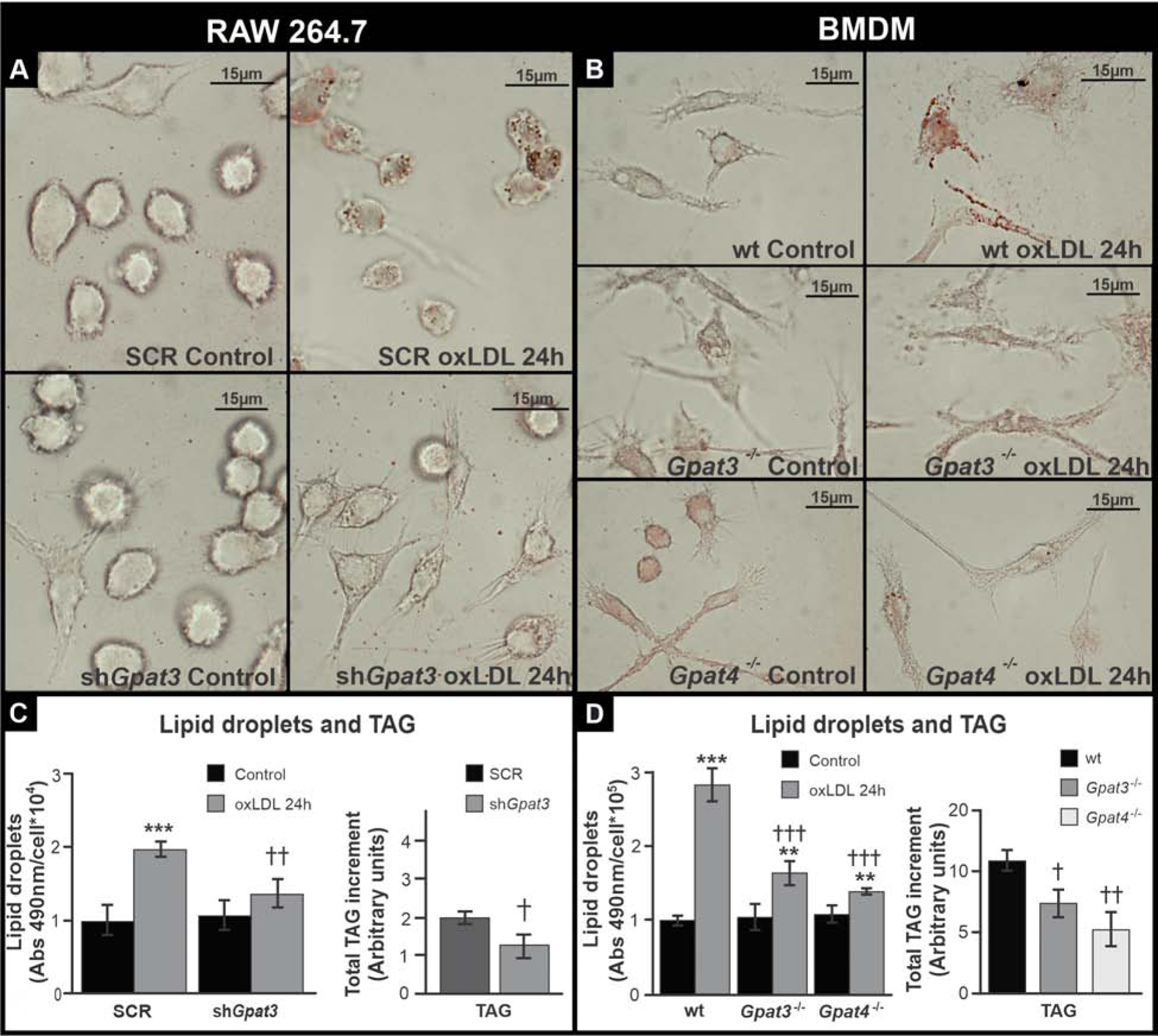
Figure 2.

Effect of GPAT3 and GPAT4 silencing or knock-out on lipid accumulation after foam cell formation.
(A and C) RAW 264.7 SCR and shGpat3 cells. (B and D) Wt, Gpat3−/− and Gpat4−/− BMDM. (A and B) Cells were treated for 24 h with oxLDL, fixed, and stained with Oil red-O to visualize LDs. (C and D) LD and TAG content was analyzed colorimetrically after 24 h oxLDL treatment. Data are from three independent experiments. *** p<0.001, ** p<0.01 with respect to controls. ††† p<0.001, †† p<0.01 and † p<0.05 with respect to wt or SCR + oxLDL.
