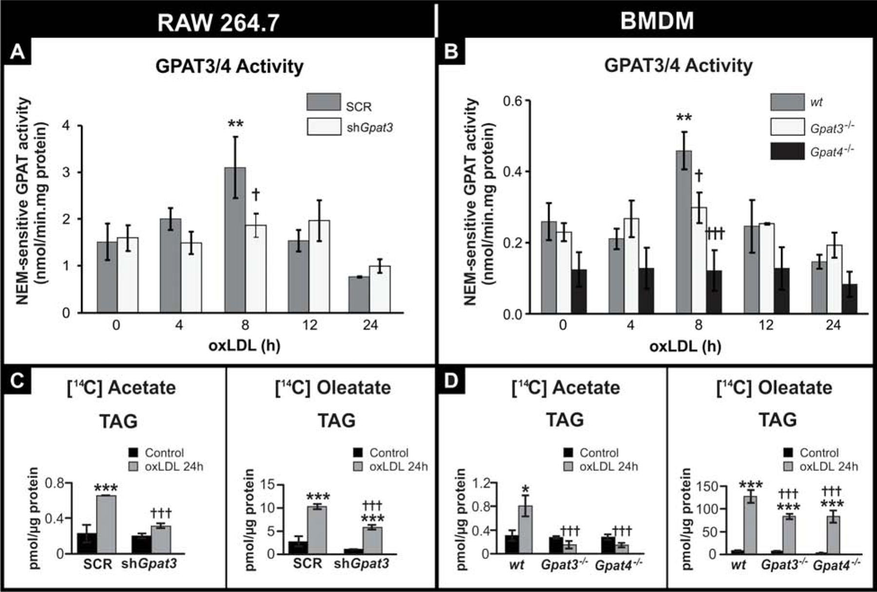
FIGURE 3.

Effect of GPAT3 and GPAT4 silencing or knockout on GPAT activity and TAG synthesis after oxLDL treatment.
(A and C) RAW 264.7 SCR and shGpat3 cells. (B and D) wt, Gpat3−/− and Gpat4−/− BMDM. (A and B) Cells were treated for the indicated times with oxLDL, and GPAT activity was assayed in total membranes in the presence or absence of NEM. NEM-sensitive GPAT activity (corresponding to GPAT3/4 activity) was calculated by subtracting NEM-resistant GPAT activity from the total. (C and D) Cells were treated for 24 h with oxLDL and 2.2 mM [14C]acetate or 0.1 mM [14C]oleate. Lipids were extracted and separated by TLC. Radiolabeled TAG were quantified with a Bioscan scanner. Data for all figures are from three independent experiments. *** p<0.001, ** p<0.01, with respect to controls. ††† p<0.001 with respect to wt or SCR + oxLDL.
