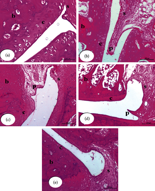
Figure 3.

Photomicrographs of (H & E 200×) stained sections of right hind leg ankle joints show histopathological effects of BM-MSCs and/or IMC treatments on CFA-induced arthritic rats. Ankle joint section from normal rat (a) shows normal structure of the synovial membrane (s), cartilage (c), and bone (b). Ankle joint section from CFA-induced arthritic rat (b) shows the substantially expanding synovial membrane with sever pannus formation (p). Ankle joint section from CFA + BM-MSCs rat (c) shows the synovial membrane with mild infiltration and pannus formation (p). Ankle joint section from CFA + IMC rat (d) shows moderate infiltration and pannus formation. Ankle joint section from CFA + BM-MSCs plus IMC rat (e) shows nearly normal histological structure of the hind ankle joint with a normal synovial membrane and mild cellular infiltration.
